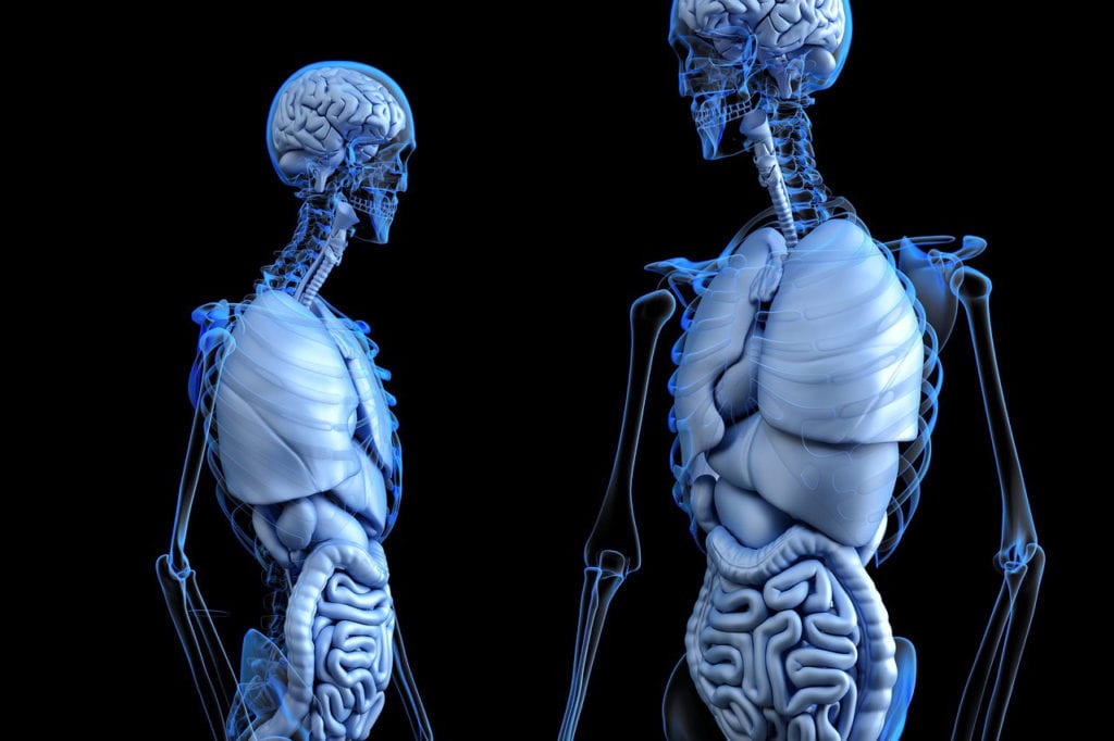
Even as we advance in science and medicine, human organs are still complicated and largely mysterious. They’re affected by lots of external factors, changing with temperature, pH, and access to nutrients. As the world outside of us changes, so does the world inside. These factors alter the populations of microorganisms, such as bacteria. Microbiomes don’t just change from one person to the next, but within each part of each person. Even in just one lung, the bacterial make up will change dramatically from one side to the next.
While we can administer treatments which alter these bacteria populations, the bacteria populations also affect the response an individual has to a treatment.
Naturally, this is a facet of medicine that has previously overwhelmed researchers. There’s so much variation between people, between organs, between different parts of one organ. There’s so much data. How do you begin to understand that landscape?
A research team at the University of California San Diego might be onto something. The team, led by Pieter Dorrestein, recently published their results. They created the first 3D visualization tool that allows researchers to map large amounts of microbe data onto a whole organ. Scientists can use this model and test out what happens when they add another chemical factor, like medicine, into the community.
Right now, the team focuses on using this model to find a targeted treatment for cystic fibrosis.
Cystic fibrosis is a rare, progressive disease that leads to digestive and respiratory damage. It’s caused by a genetic mutation that alters the way the body regulates salt. Treatment is mostly oriented at managing infections, while researchers look for a more comprehensive cure. To learn more about cystic fibrosis, click here.
The research team essentially built the 3D reconstruction from many radiological scans. They used a lung from a patient who had suffered from cystic fibrosis, and dividing it into sections. After that, they examined each section for microorganisms, their metabolites, and medication the patient had received. One of the researchers used a google chrome extension called “ili” to create the visualization that had never been seen before: an organ with its microbiome.
The model works in 2D or 3D. This means the microbiome mapped on it is not a superficial layer on the surface. The microbe populations are present, even in the internal layers of a lung.
The researchers could then apply antibiotics to the model, and literally see how the microbe population reacted. They could observe things that were previously impossible to see or predict.
The researchers were able to finally see how drugs interacted with a microbe population. For example, the patient whose lung they used had received antibiotics to manage their cystic fibrosis. Because of a bacteria population, it wasn’t able to reach the bottom of the patient’s lung.
This knowledge has significant implications regarding how we treat cystic fibrosis. However, it doesn’t stop there. The team hopes this tool will be used by other researchers, to better understand other diseases and their treatments. They’re especially interested in seeing it used for research around tumors, as they come with a unique microbiome. The visualization tool is open-source, and could change the way we understand different diseases, and the tiny world inside of us.