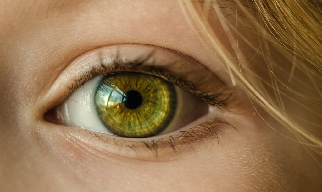
The BIOIMAGING 2018 Conference in Portugal recently announced it’s “Best Student Paper” award. The award went to a Nigerian doctoral student who identified a new technique for examining the retina. This technique improves the chances for doctors to catch diseases like glaucoma and others that damage the human retina. Keep reading to learn more about this development or follow the original story here.
Bashir Dodo studies at Brunel University London. He specializes in computer science. Dodo received the award for best student paper for a technique that will improve the diagnostic processes involving the retina.
Dodo’s work centers on an algorithm he demonstrated in his paper. The algorithm interacts with Optical Coherence Tomography (OCT) equipment used to examine the eye. In combination with OCT devices, Dodo’s algorithm allows for the creation of segmented images of the retina. The algorithm also uniquely separates this images into seven distinct layers.
Scientists hope that procedures based on Dodo’s research will be able to more accurately and efficiently provide diagnoses. Increases in speed and accuracy could save the sight of patients if damage is noticed early enough.
Dodo drew inspiration for his work from the concept of similarity in psychology. He based his OCT algorithm on the concepts of continuity and discontinuity. This allows the algorithm to distinguish when one layer of the retina transitions to the next.
As Dodo explains, layer imaging is already in clinical use. It forms one of the early steps in OCT retina image analysis. In the case of Glaucoma, for example, the thickness profile of the retinal nerve is an important piece of the diagnostic puzzle. The thickness of the this nerve fiber can be determined by calculations determined from the segment layer.
Admittedly, doctors can already identify the layers from OCT scans. Doctors must, however, perform this process manually. It takes time, and leaves potential for human error. Dodo’s novel approach segments the images of the retina automatically. This allows specialists to focus on spotting abnormalities. It quickens the process. Furthermore, the automatic segmentation allows for more efficient tracking of treatments.


