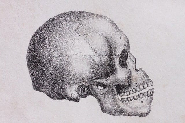
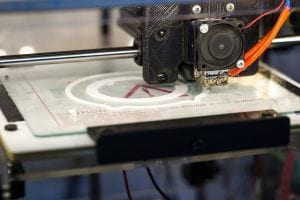
Doctors are using three-dimensional printed skulls to help prepare for surgery and as a tool to educate surgeons. With the 3-D models, surgery planning can be custom-tailored to each specific case. CAT scans are used to make a three dimensional electronic image of a patient’s skull and those scans are to translated into a three-dimensional, life-sized model. his allows surgeons to study the patient’s skull prior to surgery and be better prepared of what to expect. 3-D printed skulls will also be used in teaching cranial surgery techniques to surgeons. The development of 3-D printed skulls will be beneficial for children born with cranial birth defects, such as craniosynostosis.
Read the full article about this exciting development from UVAToday.
What are your thoughts on rare disease research? Share your stories, thoughts and hopes with the Patient Worthy community!


