In a press release on PRWeb, the Southwest Eye Institute explains how Dr. Ahmed Soliman recently performed a Bowman layer transplant to treat keratoconus. This unique and minimally-invasive procedure, which can be used as an alternative to a full corneal transplant, has only been performed at Southwest Eye Institute and one other location in the United States.
Keratoconus
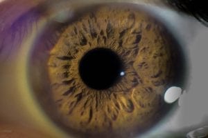
Keratoconus is a progressive eye disorder in which the cornea thins, resulting in a conical bulge in or near the middle of the eye. Doctors aren’t quite sure what causes keratoconus. However, some believe it might result from genetic or environmental causes. This disorder is common in patients with Down syndrome and Marfan syndrome.
Symptom onset usually occurs between ages 10 and 25. These symptoms can last up to, or even longer than, 10 years. Symptoms include:
- Light sensitivity
- Blurred or cloudy vision
- Progressively worsening vision
- Frequent changes to eyeglass or contact prescriptions
- Difficulty driving at night
Learn more about keratoconus.
Texas’ First Bowman Layer Transplant
The Cornea
Your cornea is the clear layer on the front of your eye. It helps allow light into your eyes so that you can properly see. Additionally, the cornea plays a role in focusing. According to All About Vision, the cornea is made up of 5 distinct layers:
- Corneal epithelium: This is the outermost layer and makes up about 10% of the cornea’s thickness. Epithelial cells are produced each week and then sloughed off when dead. Your corneal epithelium lasts about one week before new cells are produced.
- Bowman layer: The Bowman layer is a thin, but dense, sheet of connective tissue that sits between the corneal epithelium and the corneal storm.
- Corneal stroma: Overall, the corneal stroma makes up about 90% of the cornea’s thickness. The structure of connective tissue called collagen fibrils helps the cornea remain clear and focused.
- Descemet’s membrane: This membrane is pretty thin, but thickens slightly the older you get. It sits in between the corneal stroma and the corneal endothelium.
- Corneal endothelium: This is an extremely thin layer of cells that makes up the final layer of the cornea.
Bowman Layer Transplant to Treat Keratoconus
There are a few potential ways to treat keratoconus, such as hard contact lenses. But over time, these may become ineffective or uncomfortable. However, in some cases, patients need a cornea transplant to regain their vision.
How does a cornea transplant work? Surgeons remove the cornea and transplant a donor cornea. Because it requires stitches, it is considered an invasive surgical procedure. In some cases, following the procedure, the patient’s body might reject the donor cornea. However, if not, there is still a good amount of follow-up needed. In particular, patients are required to use steroidal eye drops. But using these for too long can cause other issues, like glaucoma.
What is a Bowman layer transplant and why is it such a unique option? A Bowman layer transplant is much less invasive, making it an easier and less problematic option for some patients. Prior to its use at Southwest Eye Institute, only one other location in the United States offered this procedure. Whereas a corneal transplant uses an entire donor cornea, the Bowman layer transplant uses only (you guessed it) the Bowman layer and some surrounding cells. Dr. Soliman integrated the Bowman layer into the patient’s cornea to flatten the bulge and prevent keratoconus progression. No stitches are needed. Although eye drops are required, treatment is for a shorter time.