By the time glaucoma, a group of conditions characterized by progressive optic nerve damage, manifests in symptoms, it is often too late to restore or recover lost vision. By 2040, researchers estimate that an estimated 111.8 million people across the globe will have glaucoma and, unfortunately, many cases will go undiagnosed. Screening requires certain technology and can be extremely time-consuming. According to Medical XPress, a team of doctors and scientists from Singapore are working to overcome these barriers through artificial intelligence (AI).
In fact, this team has created a novel AI screening method based on a variety of machine learning techniques to optimize the diagnostic process. By evaluating stereo fundus images, as well as certain algorithms, the screening method can decipher between glaucoma and healthy optic nerves. Interested in learning more? Take a look at the research in Methods.
Glaucoma
So what exactly is glaucoma? Glaucoma is comprised of a variety of different conditions in which the optic nerve is progressively damaged, often due to ocular pressure. However, doctors are not sure exactly why this occurs, though some hypothesize it could be due to gene mutations or eye injuries.
Normally, the optic nerve transmits images to the brain. However, when damaged, vision loss occurs. Some rare subtypes of glaucoma exist, such as congenital glaucoma and pseudoexfoliation syndrome. Symptoms do differ based on the subtype. However, some symptoms which may appear include:
- Peripheral vision loss
- Blurred vision
- “Halos” around light
- Headache
- Eye pain
Using Artificial Intelligence
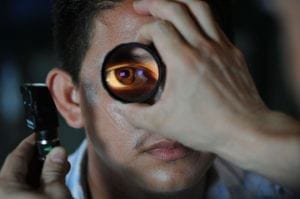
To develop their artificial intelligence screening tool, the research team created algorithms that read and analyze stereo fundus images. As described in the Journal of Ophthalmic Photography:
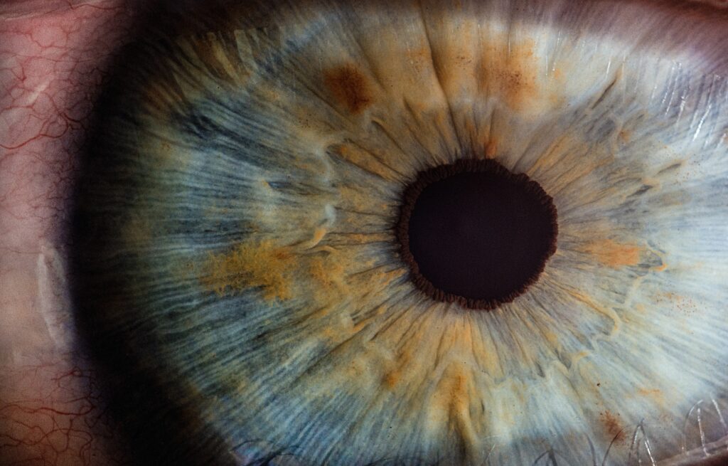
In stereo fundus photography, two images are created photographically and, when viewed, become fused in the brain. When you view the images, your left eye views the left image, your right eye views the right image, and your brain then recreates the depth relationships that were observed at the time of photography.
In short, stereo fundus photography uses multiple 2D images of the retina, from multiple angles, which combine to form a three-dimensional photo. Once the research team had these images, they ran a set of algorithms using a deep convolutional neural network and an attention-guided network. Finally, these algorithms produced predictions on whether the images suggested glaucoma. In their test, researchers used 70 fundus images from those with glaucoma and 212 fundus images from healthy individuals and trained the algorithms using 70% of the dataset. Ultimately, researchers found that:
- When applying the algorithms on the remaining 30%, the artificial intelligence screening system accurately noted 97% of glaucoma cases with 95% sensitivity. This is significantly higher than other similar tools or technologies.
- The researchers also found that using more fundus imagery increased the sensitivity.
Ultimately, researchers hope that moving forward, the tool can be used to improve glaucoma screening in underserved communities.