Welcome to Study of the Week from Patient Worthy. In this segment, we select a study we posted about from the previous week that we think is of particular interest or importance and go more in-depth. In this story we will talk about the details of the study and explain why it’s important, who will be impacted, and more.
If you read our short form research stories and find yourself wanting to learn more, you’ve come to the right place.

This week’s study is…
Estimation of visual function using deep learning from ultra-widefield fundus images of eyes with retinitis pigmentosa
We previously published about this research in a story titled “Using Deep Learning to Assess RP Disease Progression” which can be found here. The study was originally published in the research publication the JAMA Ophthalmology. You can read the full text of the study here.
This retrospective study involved five different institutions in Japan, including Saneikai Tsukazaki Hospital, Kobe City Eye Hospital, Tokushima University, Kyoto University, and Chiba University
What Happened?
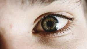
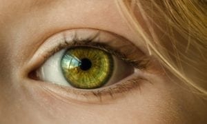
Retinitis pigmentosa is a genetic disorder that causes a decline in vision. Currently, there is no widely available effective therapy for the disease. The goal of this study was to develop a method for predicting progression of retinitis pigmentosa vision decline. Using retrospective study data, the research team sought to utilize deep learning techniques and determine if it could accurately estimate progression using ultra-widefield fundus autofluorescence images.
Deep learning is an AI network in which computers are trained to process and analyze a given set of data in order to reach a certain objective governed by an algorithm. The data for the study was collected between January 2012 and December 2018. 695 patients living with retinitis pigmentosa were included in the study data. The ultra-widefield images were used to train the algorithm and evaluate 31 ‘ensemble’ models which were derived from five distinct deep learning models.
The model that solely used the images for prediction provided the most accurate assessment of changes in vision over time, likely because the type of image used likely had the most data for the model. The researchers concluded that the deep learning model that they used, which was based on the ultra-widefield fundus autofluorescence images, has the potential to be an effective and non-invasive method of predicting disease progression in retinitis pigmentosa.
About Retinitis Pigmentosa
Retinitis pigmentosa is a genetic disorder that affects vision. This disease can progress to total blindness, although this is rare. This disorder can appear on its own or as a complication alongside other disorders or systemic illnesses. Retinitis pigmentosa is usually inherited from the patient’s parents, and a number of genetic mutations can cause it to occur. Symptoms of this disorder include generalized vision loss, difficulty adjusting to lighting conditions, aversion to bright light, night blindness, tunnel vision, and blurred vision. There is a serious lack of treatment options for the condition, and there are no therapies that can definitively delay, halt, or reverse the decline in visual acuity. A retinal prosthesis, or bionic eye, is available in some places and can help improve vision to a limited degree. Gene therapy could be a viable option in the future. To learn more about retinitis pigmentosa, click here.
Why Does it Matter?
The results from this study appear to indicate that evaluation of the visual function of people living with retinitis pigmentosa using ultra-widefield fundus autofluorescence images with deep learning could be a method for accurately and objectively assessing the progression of the disease. Deep learning models may also be able to assess progression during follow up.
The team did note some limitations of the study. Patterns of progression in the condition vary greatly, so it’s not clear if the approach can work in progression patterns that differ from those found in the dataset. The scientists also aren’t entirely certain what the model uses to estimate visual function.