Researchers have recently released some exciting news for Gaucher disease patients: a combination of two imaging techniques has been shown to better evaluate the risk of Gaucher patients developing bone fractures.
The first of these techniques was a novel method known as a trabecular bone score (TBS). The second technique of the combination was a bone mineral density technique. These were both noninvasive.
These two techniques were combined in a recent study whose findings were recently published the Blood Cells, Molecules & Diseases journal.
Gaucher disease is a rare disease that affects 1 in 40,000 live births in the general population. Basically, it is a disease that starts when a person does not have a specific enzyme in the body to break down a fatty chemical called glucocerebroside. The lack of this enzyme, called glucocerebrosidase (GCase), consequently allows fat-laden Gaucher cells to accumulate in places such as the spleen, liver and bone marrow. To learn more about Gaucher disease, click here.
At the moment, the treatment called enzyme replacement therapy (ERT) is the most used treatment technique for patients with Gaucher disease. Though this technique has improved the condition of these patients, there are some drawbacks as well.
The study’s author describes that ERT has historically been proven to be effective in improving complications from Gaucher disease, specifically with bone marrow infiltration, but the effects of ERT on bone mineral density vary and also need a lengthy treatment regimen to counteract.
Thus, it is very important for Gaucher disease patients to undergo screenings to assess the severity of bone disease in their particular condition. These assessments not only help measure the level of severity in the particular patient’s condition, but also can aid in better understanding Gaucher disease in general.
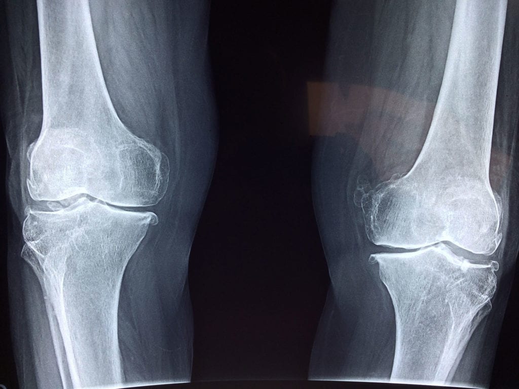
With this in mind, the authors of this study sought to test the effectiveness of new techniques (specifically by evaluating the shapes, architecture, and geometry of the bones). With these, they hoped to accurately predict the Gaucher disease patients who are at increased risks for developing bone complications.
In particular, these authors used “noninvasive DXA-based techniques: TBS and Hip Structural Analysis (HSA).” These two techniques were used on 23 Gaucher disease patients, and in conclusion, the study showed that these techniques were useful in diagnosing the skeletal aspects of Gaucher disease.
These patients, in particular, had a heightened risk of bone fractures and cell death in bones, or “avascular osteonecrosis,” according to the study. Thus, having new resources to assess these complications is great news for the Gaucher community.
Hopefully, the results from this study can lead to not only imaging improvements but also treatment advancements. To read more about this study, click here.
What are your thoughts about ongoing research for rare diseases? Share your thoughts, and your hopes, with the Patient Worthy community!