For many years, scientists have known about toxic clumps forming in patients with Huntington’s disease (HD). However, until recently, scientists relied on examining post mortem brain samples of HD animal models, or samples from the brains of patients who donated their brains for research.
According to a recent article published in HDBUZZ, scientists from Italy, Germany, the UK, Sweden and the US have created molecular tools (scanners) that allow them to “see” the toxic clumps in animals. This international group is also developing additional versions of the scanner as a backup. The next step will be scanning the clumps in humans.
About Huntington’s Disease
HD is defined as an acute disturbance of the central nervous system caused by a faulty gene that produces a defective protein (the Huntingtin protein). The protein accumulates in the body as HD progresses and attacks clusters of nerve tissue(neurons) within the brain that control coordination.
Humans have two copies of the normal huntingtin gene. In HD, however, one of the genes is mutated and contains a section of DNA code that is, for reasons yet unknown, copied over and over. This is known as repeat expansion.
The disease is characterized by involuntary movement of muscles and intellectual deterioration.
About the PET Tracer
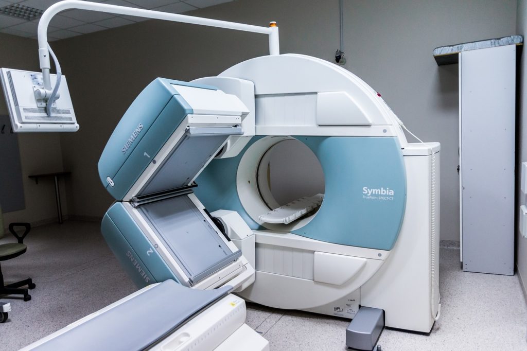
The new molecular tools which bind to the protein clumps have radiolabels causing them to light up under a positron emission tomography (PET) scan. They are called PET Tracers.
Various types of tracers may be used in diagnostic settings. They may be in the form of injections or they may be inhaled or swallowed.
When the tracer is positioned within the body and the target has been located, that part of the body will light up.
One advantage of the new method is that the PET scan can be used many times within a patient’s lifetime.
Another advantage is that the PET scans allow the doctors to look at the brain as opposed to using invasive procedures limiting the doctors to simply measuring spinal fluid.
And lastly, since the clumps are the result of a form of mutant huntingtin protein, researchers will be able to measure any changes to that toxic form. The current method of measurement is unsatisfactory as it measures all forms of huntingtin, even the healthy protein.
Looking Forward
A clinical trial will soon investigate the tracer in humans. PET/MRI will be used to observe how PET follows huntingtin’s progress within the brain. Clumps in an HD patient’s brain can be used as biomarkers which are reference points in many diseases.
In the new scans, they are measurements that scientists use to keep track of HD’s progression.
The new tools are a first-time event. This is the first time scientists can track the Huntingtin protein throughout the brain of HD patients.


