In the past, there have been a small number of studies exploring the connection between sickle cell disease (SCD) and craniosynostosis. More recently, researchers sought to understand whether there was an actual association between the two conditions. According to Rare Disease Advisor, a research team performed a retrospective review and found that there was a significant prevalence of craniosynostosis in those with SCD.
What are Craniosynostosis and Sickle Cell Disease?
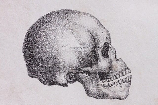
Craniosynostosis is a birth defect and disorder in which one or more fibrous joints (sutures) between the bones of an infant’s skull close prematurely. At this point, the skull has not been fully formed. Despite this, the brain continues to grow. As a result, craniosynostosis may cause a misshapen head appearance. The exact cause is unknown, though there is a suspected relationship with genetic disorders like Pfeiffer syndrome. Symptoms and characteristics include a misshapen skull, an abnormal or disappearing fontanel, a hard raised ridge along affected sutures, slowed head growth, irritability, excessive sleepiness, poor feeding, a high-pitched cry, and projectile vomiting.
Sickle cell disease (SCD) refers to a group of rare genetic disorders characterized by malformed, sickle-shaped blood cells. SCD results from mutations on the hemoglobin-Beta gene, which is found on chromosome 11. Normally, hemoglobin carries oxygen throughout the body. But in SCD, the sickle-shaped cells block and restrict blood flow, causing symptoms and other health issues. Symptoms can include pain crisis, jaundice, swollen hands and feet, anemia, delayed growth, fatigue, and organ damage.
Unpacking the Research
Within their retrospective review, the research team analyzed 94 CT scans of the head in pediatric patients with SCD between ages 0-8. These CT scans were taken over a 10-year period and were not related to head shape abnormalities. A majority of the patients were male and a majority were African-American. 80.9% had homozygous SCD, 11.7% had heterozygous SCD, and 7.4% had beta-thalassemia.
In the findings, published in Plastic Surgery and Reconstruction – Global Open, the researchers explained that 19.1% of patients had craniosynostosis. Additional findings included:
- Despite a majority of study participants being male, 55.6% of those with craniosynostosis were female. All those with craniosynostosis were African-American.
- There are several types of craniosynostosis based on which suture fused prematurely (sagittal, coronal, metopic, lambdoid). In this case, all craniosynostosis cases were the result of early sagittal suture fusion.
- Those with craniosynostosis were more likely to have a family history of neurodevelopmental disease or SCD, and were more likely to have been treated with folic acid supplements. In fact, having a first-degree relative with SCD increased the risk of developing craniosynostosis by 14-fold.
- Those with SCD-associated vasculopathy were 8x more likely to develop craniosynostosis.
Ultimately, the researchers explain that more research is needed to better understand the relevance of their findings. However, they also share the need for increased awareness – and advocate for doctors whose patients have SCD to be prepared to potentially identify craniosynostosis.