The European Society of Cataract & Refractive Surgeons (ESCRS) held its ESCRS Winter Meeting from February 15-18, 2024. During the meeting, Dr. M.S. Swathi, MD, shared data from a pilot study that sought out to learn how intrastromal lamellar keratoplasty with collagen cross-linking biomechanically changed the cornea in people with progressive keratoconus.
Before we dive into the results, let’s first look at what these two procedures are. An article published in the Journal of Cataract & Refractive Surgery, authors describe the process of an intrastromal lamellar keratoplasty:
After a pocket is created along the marked margins of the thinned inferior cornea with Melles dissectors, a crescent of the same size is marked on the donor cornea. The epithelial and endothelial layers are removed, and the donor tissue is cut in a crescentic shape in the size of the marked corneal pocket. The donor crescentic stroma is then inserted into the stromal pocket.
This helps to reinforce the thin portion of the cornea. Collagen cross-linking, which is also known as corneal cross-linking, is described by the American Academy of Opthalmology as a surgery to fix a weakened cornea:
Disease or sometimes surgery can harm collagen, an important substance that holds the cornea together. By “cross-linking” new collagen fibers together, CXL surgery strengthens and reinforces the cornea.
Biomechanical Changes in Keratoconus
According to Michela Cimberle in Healio, Dr. Swathi explained how her team wanted to understand what biomechanical changes occurred in the cornea following intrastromal lamellar keratoplasty with collagen cross-linking. The study included 10 people with progressive keratoconus with a six-month follow-up period.
To begin, the patients underwent intrastromal lamellar keratoplasty to include a donor lenticule. The doctors then removed the epithelium before continuing with collagen cross-linking. Over the follow-up period, the researchers noted that:
- Both uncorrected and best corrected visual acuity improved by ~1 line.
- The corneas flattened and also improved in thickness, up to 428-557 µm.
- Following the procedures, the corneas became stronger and more stable.
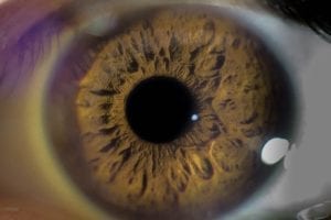
About Keratoconus
Keratoconus is an ocular (eye) disorder that affects the cornea. This disorder is characterized by progressive (worsening) corneal thinning and a conical bulge near the center of the eye. Doctors don’t know what causes keratoconus. Right now, the hypothesis is that there are a mix of genetic and environmental factors at play. Keratoconus is more common in people who have Marfan syndrome, Leber congenital amaurosis, and Down syndrome. This condition typically affects both eyes. However, it may progress differently. Symptoms, which usually begin between 10 to 25 years old, may include:
- Blurred or distorted vision
- Photophobia (increased light sensitivity)
- Poor night vision
- Double vision
- Nearsightedness
- Loss of visual acuity
Treatments for keratoconus include corrective eyeglasses or lenses, corneal transplants, corneal cross-linking, and surgery. If you have keratoconus, please speak with your doctor about the best course of treatment.