Christina (“Chris”) Coates has lived a full, joyous, and fulfilling life. The Arizona native has built something beautiful: a family with two daughters, two cats, two dogs, and a loving husband. After receiving her degree in accounting, Chris began working in credit and finance for a heavy-duty machinery company, a job that she has thrived in for the last sixteen years (although she is now on medical leave). Ultimately, she shares:
“I had a very boring life—a great one, don’t get me wrong, but a boring one—until I was about 40 years old, and then all the wheels started falling off.”
It was around this time that Chris’ health situation began to change. After undergoing brain surgery, she experienced a number of worrying symptoms. Further testing eventually found that Chris was living with hypertrophic olivary degeneration (HOD), a rare neurological condition.

Since her diagnosis, Chris has become a fierce advocate for awareness and research. She has formed the Hypertrophic Olivary Degeneration Association (HODA) and provided a community for others facing HOD and other rare or chronic illnesses.
Recently, Chris spoke with Patient Worthy to share her story in an effort to further our understanding of what hypertrophic olivary degeneration is and offer avenues of support for others within this community.
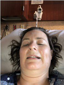
Chris’ Story
In 2017, Chris was diagnosed with a small cavernous malformation with a previous cerebellar hemorrhage. The cavernous malformation was located on the 4th ventricle of her brain (brainstem adjacent). Cavernous malformations are clusters of abnormal blood vessels with thin walls, so they can ooze or seep blood. As Chris explains:
“They can explode or hemorrhage, which can be dangerous. Because of its location, and especially because of its potential impact on cerebrospinal fluid regulation, my doctors and I wanted to remove it.”
In 2021, Chris underwent a craniotomy (a type of brain surgery) to remove the malformation. Her recovery went smoothly. Within six weeks, she was back at work. Then her vision began to change. She had significant pain in the right eye between the eye socket and eyeball, as well as nystagmus (involuntary eye movements) and blurred vision in the left eye. She shares:
“It ached and my vision was bouncing around a lot. When I spoke to my doctor, I explained that it felt like my eyes were disconnected from my brain. I was told initially that it could be an issue with my optic nerve. My neurosurgeon sent me for an MRI, which is when they found a lesion on my medulla.”
At this point, Chris’ doctors hypothesized that she had multiple sclerosis (MS). She was sent back to her neurologist, who set her up for a spinal tap and brain MRI. A multiple sclerosis diagnosis requires two lesions and a spinal tap.
Yet something didn’t sit quite right with Chris. Frustrated and still in pain, she reached out to the Mayo Clinic with her medical information and reports. She was looking for a second opinion. Shortly after meeting with a Mayo neurologist who specializes in MS and more testing, a radiologist from the Mayo Clinic noted that the most recent MRI showed a lesion that resembled HOD. The Mayo neurologist reached out to Chris with their diagnosis: hypertrophic olivary degeneration. Says Chris:
“When they called me with the diagnosis, I was told that they didn’t have many cases of HOD and couldn’t give me much information about what might happen. They said that it wasn’t a death sentence, and it would resolve on its own.”
What is Hypertrophic Olivary Degeneration (HOD)?
In an article from the National Organization for Rare Disorders (NORD), using information from the National Institutes of Health (NIH) Genetic and Rare Diseases Information Center (GARD), hypertrophic olivary degeneration is described as:
“a rare neurological condition caused by degeneration in the brain stem, the structure that connects the brain to the spinal cord. HOD is considered unique because the inferior olivary nucleus (“olive”) initially becomes hypertrophic rather than atrophic (wasted) though over time, the olive goes through atrophy.”
Chris continues:
“There are two inferior olivary nuclei: the left and the right. HOD can happen in one or both of them. The olives are blown up like balloons and then they atrophy. The brainstem is the center of our most primitive actions so when you have disease there, it gets a little tricky. HOD can affect your balance, vision, the way you walk, the way you swallow. Truthfully, the condition is poorly understood. It doesn’t have an ICD-10 code so it isn’t being tracked and many patients note that their neurologists don’t tell them that they have HOD; they just find it in their reports. So we have no idea how many people are living with HOD.”
Currently, there are a known 100-150 people worldwide living with HOD. But as suggested above, it is very possible that HOD is under and mis-diagnosed.
Chris has found that HOD affects every patient differently. So far, symptoms of HOD have been found to include:
- Nystagmus
- Double vision
- Fatigue
- Myoclonus
- Headache
- Pins and needles sensations that don’t resolve quickly
- Difficulty swallowing
- Acquired ataxia
- Tremors
- Muscle weakness
- Impaired gait
Chris shares:
“I can ‘pass’ for a normal person, but you can tell that I have something going on when I get tired. It can be frustrating. People tell me that I look great. They don’t understand what it takes for me to do that just so people won’t stare. Having HOD is like being diagnosed with cancer but in the year 1650. There’s not a lot of research or information, but I want people to recognize that lived experience is still valid and should be used to shape the future medical understanding of HOD.”
Hypertrophic olivary degeneration is diagnosed following a lesion being found on an MRI. The lesion typically looks as though it is demyelinating. Most patients are under the main care of a neurologist.
Join us in Part 2 as we discuss rare disease myths, forming the Hypertrophic Olivary Degeneration Association (HODA), and advice for others.


