According to BioTechniques, American researchers have taken a new, hands-on approach to learning more about cytomegalovirus-induced microcephaly. Although cytomegalovirus has been linked to microcephaly, the topic is somewhat under researched. However, by testing and collecting data on 3D “mini-brains,” researchers can better understand how cytomegalovirus can contribute to congenital defects.
View their findings in Cell Reports Medicine.
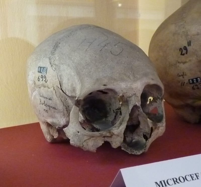
Microcephaly
Microcephaly is a rare neurological condition characterized by an extremely small head at birth. Basically, babies born with microcephaly did not fully develop their brains or damaged their brains within the womb. It can occur alone or in conjunction with other birth defects. Causes of microcephaly include genetic mutations, malnutrition, toxicity exposure, craniosynostosis, and infections during pregnancy.
Symptoms can be mild to severe. In addition to a smaller head, symptoms of microcephaly include vision and balance problems, hearing loss, developmental and intellectual delays, seizures, and facial distortions. Learn more about microcephaly.
Cytomegalovirus (CMV)
As previously stated, infections during pregnancy can lead to microcephaly. One common viral infection is cytomegalovirus. According to the CDC, around 33% of children contract cytomegalovirus before age 5, and around 50% of people have it by the time they turn 40. It transmits through bodily fluids. Symptoms often include fever, swollen glands, fatigue, and a sore throat. However, most people will never know they have CMV because of a lack of symptoms or impact.
But while cytomegalovirus is not a threat to the greater public, it can cause complications during pregnancy. Around 20% of babies infected with CMV experience birth defects or other health problems.
3D “Mini-Brains”
Prior research on the intersection of CMV and microcephaly has been shaky at best. This is because, as a neurological disorder, the impact of microcephaly on 2D or animal models is difficult to replicate. So, researchers in California took it a step further: 3D organoid “mini brains.”
According to a Nature article, organoids are:
3D multicellular in vitro tissue construct[s] that mimic its corresponding in vivo organ, such that it can be used to study aspects of that organ in the tissue culture dish…the word organoid is most commonly used to describe such constructs derived from stem cells.
In vitro means cells or tissue created or analyzed outside of a living organism. In vivo means within a living organism. So, brain organoids are similar to to actual brain tissue, but without scientists needing to actually source a brain for study. In this case, researchers used mini-brains to model how CMV can cause brains to shrink.
After injecting the mini brains with a specific CMV strain (TB40/E), researchers saw a significant reduction in brain growth. Additionally, the brains lacked structure and neuronal function.
Next, researchers tested a CMV-induced microcephaly vaccine on the mini brains. They found that the neutralizing antibodies (NAbs) helped protect the tissue against further damage. Now, researchers believe that early detection and treatment may prevent or reduce the impact of microcephaly. However, real-world studies are needed to determine replicability.