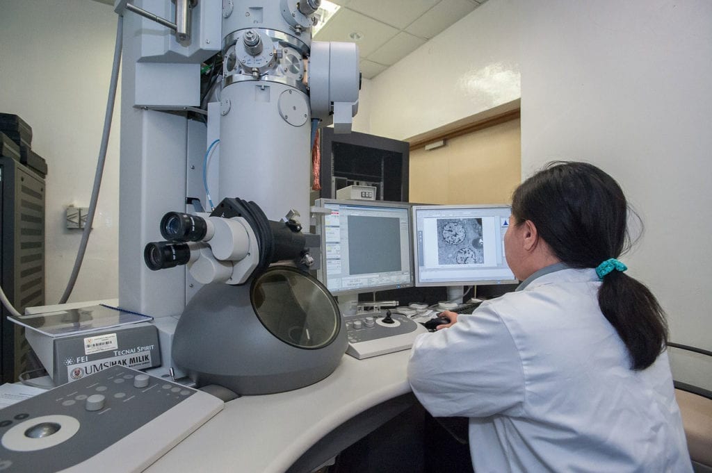
According to a story from EurekAlert!, a collaborative team of researchers from the University of Florida and The Salk Institute are using cryo-electron microscopy in order to more closely examine the structure of the AAV2 virus. This type of virus has played a role as a delivery mechanism for gene therapies in the past, and the scientists are using the this innovative imaging technology to learn more about AAV2 in order to make it even more effective in the future.
New Innovations, Better Results
This information about the structure of the virus is the result of the team’s willingness to experiment with theoretical procedures in order to enhance the capabilities of cyro-electron imaging. These procedures have allowed for the creation of a 3D model of AAV2 with higher resolution and overall detail than ever before. This new information about the structure of this virus can be utilized to develop improved gene therapies that can treat a variety of rare diseases such as hemophilia, sickle cell anemia, Leber congenital amaurosis, Duchenne muscular dystrophy, and junctional epidermolysis bullosa.
About The Study
The research focused on a variant of AAV2 which is uniquely suited for use in gene therapies. This type has an unusual modification to its amino acids, and it is also less infectious than other types of AAV2. The research also focuses most specifically on modifications to a portal which is essential for the packaging of genetic material. One of the challenges of using viruses for delivering gene therapy is that the body can recognize them as potential threats and destroy the viruses. This research could help minimize this issue.
AAV2 was also an ideal candidate for study using cryo-electron microscopy because if its high degree of symmetry. The team was able to get 60 times the data that they normally would, and this data is expected to be highly relevant to future research.
Future Research
The new procedures also allowed for data to be obtained using lower voltage microscopes; cryo-electron microscopes are prohibitively expensive for some institutions, but newer, cheaper models will soon become available, and the procedures pioneered in this study will enhance their effectiveness.
Hopefully, continued imaging studies will be able to fine tune viral vector gene therapies even further. To check out the original study, click here.