Saarland University’s medical team, led by Professor Bergita Ganse, has discovered an entirely new approach for monitoring the healing of bone fractures through measuring blood supply to the tissue at the site of the fracture, and the oxygen levels in the blood.
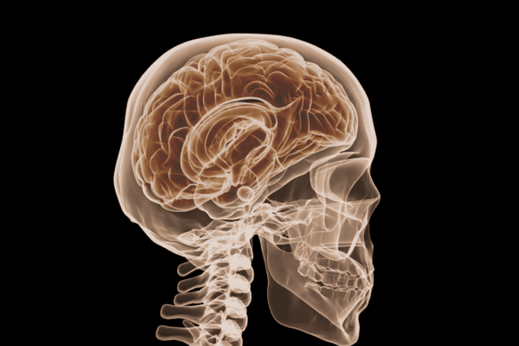
To further clarify, it is now possible to monitor bone regeneration by using near-infrared light instead of harmful (shorter) wavelength radiation. Currently, doctors are relying upon x-ray images and CT scans providing an occasional snapshot of the fracture site to monitor recovery.
Note that the findings of Prof. Ganse and her associates have been published in Biosensors and Bioelectronics and The Journal of Functional Biometrics.
About the Concept
The concept involves a commercially available pocket-sized device that is placed on the skin just above the fracture site; within seconds doctors can see how well the fracture is healing. If the patient is wearing a plaster cast, a small opening is made into the cast to allow skin contact. Therefore, the new process allows monitoring of blood oxygen and blood flow without subjecting the patient to harmful short-wave radiation.
Professor Ganse explained that their process uses devices for the examination of blood flow and saturation of oxygen in muscles that reach the bone tissue. Bone fractures have, until recently, been monitored through X-ray imaging or computed tomography scans. This exposes the patient to radiation that should not be repeated too often.
Delayed sensitivity to early healing activity is another drawback. As the fracture heals the fracture gap is covered with soft bone tissue. However, at this the bone density is not high enough to be detected by X-rays. Bone density is not sufficient until, late in the healing process. This additional monitoring of bone healing provides an improved understanding of the healing process and may detect potential complications earlier.
It has been established through the study of lower leg fractures that complications will arise in fourteen out of every one hundred cases. As Prof. Ganse explained, the sooner the doctors notice that something has not been progressing as expected, the earlier the staff can intervene.
The Complexities of Bone Fracture Repair
Fracture repair is a multi-phase and complex process whereby:
- The fracture is initially bridged with a thin covering
- In time, new bone tissue grows and is supplied with blood along with the formation of new blood vessels.
Note that to date, one problem with the new light-based method of monitoring is that the researchers are unable to probe fractures that are positioned over five centimeters under the skin.
The Professor’s work is part of a “Smart Implants” project that was launched five years ago. It is an interdisciplinary project involving various research groups at the Saarland University in the fields of medicine, computer science and engineering. Thus far the teams have developed several prototypes that provide micromechanical stimulation of the fracture site.
Prof. Ganse collaborates with NASA in the U.S., the German Aerospace Center and the European Space Agency studying how muscle and bone degrade in space. The Professor’s research has contributed to training programs geared to assisting astronauts in counteracting musculoskeletal degeneration during protracted space missions.