
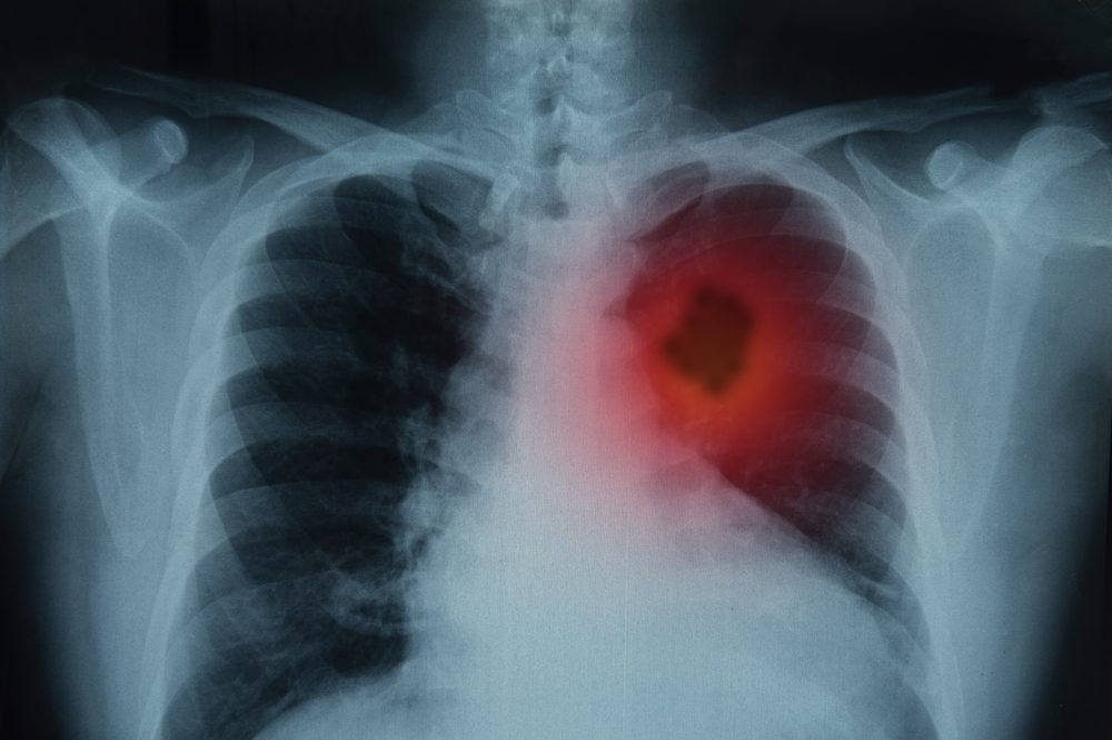
As reported on Inside Precision Medicine, scientists have developed a way to spot a specific lung cancer mutation in just minutes instead of weeks, using a combination of advanced laser imaging and artificial intelligence.
How the Technology Works
Currently, when a patient is diagnosed with lung cancer, doctors need to know if the tumor has a mutation in a gene called EGFR. This gene controls cell growth, and knowing if it is mutated is crucial because it decides which drugs will work best.
To find this mutation, scientists traditionally have to run expensive, slow genetic tests that destroy the tissue sample in the process.
This new method changes that by using two tools:
- FLIM (Fluorescence Lifetime Imaging Microscopy): Every cell in our body naturally glows slightly when hit by a specific laser. FLIM measures how long that glow lasts, creating a unique “optical fingerprint” of the cell’s metabolic activity.
- Deep Learning (AI): Researchers trained an AI model on these laser scans. The AI learned to recognize the subtle “fingerprints” of mutated cells, achieving an incredible 96.6% accuracy in identifying EGFR mutations.
How This Directly Helps Lung Cancer Patients
For anyone facing a lung cancer diagnosis, this technology could be a massive game-changer in three main ways:
- Much Faster Answers (Minutes vs. Weeks)
Waiting for genetic test results is one of the most stressful parts of a cancer diagnosis. Right now, next-generation sequencing can take weeks. This laser-and-AI method can produce results in a fraction of that time. As co-lead author Dr. Ahsan Akram explains:
“Current pathways require specialized labs and next generation sequencing. These are time consuming and costly. It can take weeks to have an answer. Here, as we are taking an image using laser light the process can take minutes.”
Getting answers in minutes means patients can start the right treatment almost immediately, saving precious time.
- Saves Rare and Valuable Tissue Samples
During a lung biopsy, doctors only get a tiny sliver of tissue. Traditional genetic testing requires staining and chemically processing the tissue, which destroys it. If doctors run out of tissue, the patient might have to go through another invasive biopsy.
Because this new method only shines a light on the sample, it is entirely harmless. Dr. Akram notes:
“As we are shining laser light on the sample, without any stains, this is a completely nondestructive process. The tissue sample is intact following the image and then can be used for whatever else is needed.”
- Highly Personalized Treatment Plans
Not all EGFR mutations are the same. The AI was actually smart enough to tell the difference between the two most common subtypes of this mutation (called exon 19 and exon 21). Because these two subtypes respond differently to drugs and have different outlooks, knowing exactly which one a patient has allows doctors to tailor the treatment plan perfectly to their specific cancer.
What’s Next?
While this is a highly successful “proof of concept,” the technology isn’t in hospitals just yet. The next step is to test it on a much larger, more diverse group of patients in real-time hospital workflows to make sure it is ready for everyday medical use.